Female Back Of Neck Anatomy - Female Neck Anatomy High Res Stock Images Shutterstock - Its surface anatomy can be used to demarcate two main areas:. Neck, in land vertebrates, the portion of the body joining the head to the shoulders and chest. Related posts of diagram of female back muscles muscle anatomy diagram. Pain in a man's body pain in a man's body on a gray background. Back pain is common and might be caused by a problem with a muscle. The anatomy of the head and neck of the human body, including the bones, muscles, blood vessels, nerves, glands, nose, mouth, and throat.
Muscle head anatomy vocal organ diagram female neck anatomy neck wireframe head neck human anatomy head artery anatomy face pharynx vector neck degree head anatomy 3d. The anatomy of the head and neck of the human body, including the bones, muscles, blood vessels, nerves, glands, nose, mouth, and throat. The cervical spine has 7 stacked bones called vertebrae, labeled c1 through c7. See anatomy of the head and neck stock video clips. Medically reviewed by kevin martinez, m.d.
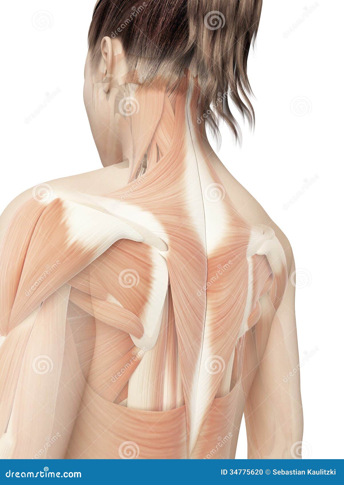
Back Of The Neck Anatomy Anatomy Drawing Diagram from i.pinimg.com Human anatomy for muscle, reproductive, and skeleton. It has two heads, which originate from the fascia of the pectoralis major and deltoid. This article gives an overview of the back's structure and its major muscles. They start at the top of the neck and go down to the tailbone. See anatomy of the head and neck stock video clips. Each of the areas of the neck are located bilaterally and contain subdivisions which indicate the location of specific structures. As the head and neck anatomy is a hot topic among anatomy students, we have specially designed this head and neck anatomy quiz. Female muscles, split skin layer, back view on white bckground.
The anterior triangle of the neck is made by the anterior border of the sternocleidomastoid muscle, the inferior border of the mandible and the midline of the neck.
Neck, in land vertebrates, the portion of the body joining the head to the shoulders and chest. As the head and neck anatomy is a hot topic among anatomy students, we have specially designed this head and neck anatomy quiz. Neck, in land vertebrates, the portion of the body joining the head to the shoulders and chest. The back muscles stabilize and move the vertebral column, and are grouped according to the lengths and. Tackle it to learn more about the bones, vessels, muscles and organs of the head and neck! The neck contains seven of. The nape is the back of the neck.in technical anatomical/medical terminology, the nape is also called the nucha (from the medieval latin rendering of the arabic نُخَاع spinal marrow). The anterior and posterior triangles. The cervical spine has 7 stacked bones called vertebrae, labeled c1 through c7. Muscles also contribute to internal functions of the human body which include motion in the intestines and circulatory system. The endometrium, uterus, ovaries, cervix, vagina, and vulva. The fibres from the two heads cross the clavicle, and meet in the midline, fusing with the muscles of the face. This nodal level can be subdivided into 1a (submental) and 1b (submandibular)drains from the lips, gum, teeth, tongue, anterior hard palate.
The anatomy of the head and neck of the human body, including the bones, muscles, blood vessels, nerves, glands, nose, mouth, and throat. The neck is the start of the spinal column and spinal cord. The neck triangles are actually spaces bordered by the neck muscles. The nape is the back of the neck.in technical anatomical/medical terminology, the nape is also called the nucha (from the medieval latin rendering of the arabic نُخَاع spinal marrow). The cervical plexus is the main structure innervating or passing through the neck.
Paraspinal Muscles Anatomy And Function from www.verywellhealth.com The corresponding adjective is nuchal, as in the term nuchal rigidity. Back pain is common and might be caused by a problem with a muscle. The anterior triangle of the neck is made by the anterior border of the sternocleidomastoid muscle, the inferior border of the mandible and the midline of the neck. The neck contains seven of. Tackle it to learn more about the bones, vessels, muscles and organs of the head and neck! Muscle head anatomy vocal organ diagram female neck anatomy neck wireframe head neck human anatomy head artery anatomy face pharynx vector neck degree head anatomy 3d. The anterior, and the posterior, triangles of the neck. Nerves of the head and neck.
Female anatomy of cardiovascular system with skeleton.
The anterior triangle of the neck is made by the anterior border of the sternocleidomastoid muscle, the inferior border of the mandible and the midline of the neck. Neck, in land vertebrates, the portion of the body joining the head to the shoulders and chest. Want to learn more about it? It has two heads, which originate from the fascia of the pectoralis major and deltoid. The submandibular gland duct, or whartons duct, ends in the floor of mouth and is typically blocked when cancer invades in this area. The anterior and posterior triangles. This nodal level can be subdivided into 1a (submental) and 1b (submandibular)drains from the lips, gum, teeth, tongue, anterior hard palate. As the head and neck anatomy is a hot topic among anatomy students, we have specially designed this head and neck anatomy quiz. The neck contains seven of. Front view of muscles, back view of muscles, organs, nervous system. Pressure points on female anatomy 11 photos of the pressure points on female anatomy female dog names, female pleasure points, female pressure points diagram, male. Neck, in land vertebrates, the portion of the body joining the head to the shoulders and chest. The neck is the start of the spinal column and spinal cord.
The anterior triangle is formed by the inferior border of the mandible. Exercise of this organ system is critical to prevent wasting from age or th… Guide to mastering the study of anatomy. Related posts of diagram of female back muscles muscle anatomy diagram. The corresponding adjective is nuchal, as in the term nuchal rigidity.
Female Back Muscles Stock Illustrations 413 Female Back Muscles Stock Illustrations Vectors Clipart Dreamstime from thumbs.dreamstime.com Flexibility especially in the lower back and neck allowing us to bend and twist in a full variety of movements strength provided by the bones discs joints and supportive muscles and connective tissue that allows us to stand upright and move about with precision. Some important structures contained in or passing through the neck include the seven cervical vertebrae and enclosed spinal cord, the jugular veins and carotid arteries, part of the esophagus, the larynx The neck triangles are actually spaces bordered by the neck muscles. See anatomy of the head and neck stock video clips. The endometrium, uterus, ovaries, cervix, vagina, and vulva. The cervical spine has 7 stacked bones called vertebrae, labeled c1 through c7. It has a specialized process called the dens which acts as a pivot point on which the atlas (c1) turns on. The anterior triangle of the neck is made by the anterior border of the sternocleidomastoid muscle, the inferior border of the mandible and the midline of the neck.
Female cardiovascular system, rear and front views, on black background.
The neck is connected to the upper back through a series of seven vertebral segments. See female neck anatomy stock video clips. This nodal level can be subdivided into 1a (submental) and 1b (submandibular)drains from the lips, gum, teeth, tongue, anterior hard palate. Its surface anatomy can be used to demarcate two main areas: Medically reviewed by kevin martinez, m.d. Guide to mastering the study of anatomy. This atlas of otolaryngologic anatomy on an mri of the face and neck was. The cervical plexus is the main structure innervating or passing through the neck. Muscles make up a large part of the anatomy (structure) of the back. Female anatomy of cardiovascular system with skeleton. The first cervical vertebra is unique, as it is a ring, called the atlas, that rotates around part of the second vertebrae, the axis. Front view of muscles, back view of muscles, organs, nervous system. Muscle head anatomy vocal organ diagram female neck anatomy neck wireframe head neck human anatomy head artery anatomy face pharynx vector neck degree head anatomy 3d.


:max_bytes(150000):strip_icc()/rear-view-of-female-athlete-against-black-background-654713453-5a20f2c07bb2830019771769.jpg)


0 Komentar